|
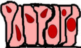
This picture shows sections through 4 different organs: Intestines, Skin, Lung, & Trachea. Note that each organ is made of multiple tissues and that their are variations on how the tissues are designed. You will learn more specific categories of these tissues . |

|
The 4 basic tissue types are Epithelium, Connective Tissue, Nervous Tissue, and Muscle Each types contains subtypes that may look different but share similar characteristics.
Below are a few examples of different tissues at high power. Under each basic category there are numerous subtypes of different looking tissue.(Rest your mouse on each picture to see a brief description of the slide) |
|||
| Epithelium | Connective Tissue | Nervous Tissue | Muscle |

|

|

|

|

|

|

|

|

|
 |

|
 |
The first step in finding epithelium on a slide is to look for free surfaces where epithelium may be present. A defining characteristics of epithelia is that they have a free, orapical, surface and a they have a basal surface. Epithelia line hollow organs (e.g., intestines, blood vessels, etc.) and cavities (e.g., abdominal cavity or pleural cavity), cover our body's surface in our skin, and make up many small tubes and ducts in the body (e.g., capillaries and ducts in sweat and salivary glands).
The free surface is what faces the opening in the organ or tube (often these are called lumens) or what faces the outside as in skin. In the body the free surface will be facing a space filled with gasses or liquid, but on a slide you typically won't see the fluid and there will be a lot more spaces than would really exist. Take the following picture for example
| On this slide of the intestines at low power, it appears that there are large areas of free surface on the left and right sides of the tissue. Are they both free surfaces? No. On the slide they are, but if this was still in a person only one of these sides faces a free surface in the lumen. In this example it is the right side of the slide. The left side is smooth muscle which NEVER lines a free surface. More examples are below. |

|
| Sometimes a slide will look like it has a lot of free surfaces to choose from. However, a free surface on a slide does not mean that there is epithelium there covering it. In this slide, epithelium does not cover any of the large visible free surfaces on the slide. This is a section of muscle and tendon that has been stuck on a slide. |
|
Once you have mastered finding appropriate free surfaces, the next step in learning the different epithelia is to determine the number of cell layers in the epithelium.
If the epithelium is only one cell-layer thick, then it is called simple epithelium and if it is more than one layer thick, then it is called stratified epithelium.
| Simple Epithelium | Stratified Epithelium |
|
|

 |
|
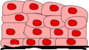
Finally, it is important to determine the shape of the apical cells (the cells at the free surface). There are three major categories for describing the shape of most epithelial cells (note most in this statement -- there are a couple of other types). These categories are squamous(flat looking cells), cuboidal (they look like cubes), and columnar (they look like columns).
Squamous Epithelium |
Cuboidal Epithelium |
Columnar Epithelium |
|
|
Do they ever look that nice and obvious on a slide? No. They usually look less defined. (Rest your mouse on each image to see what type of tissue it is)
|

|

|

|

|
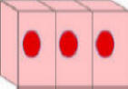
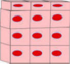
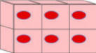
When naming epithelia, we combine the name for the number of cell layers (simple or stratified) with the shape of the surface cells (squamous, cuboidal, or columnar). For example: simple squamous epithelium, or stratified columnar epithelium, etc. There are some cartoon examples below.
What would you name this type of epithelium?
|
|
What is the name for this epithelium? | Example |
|
|
What is the name for this epithelium? | |
|
|
What is the name for this epithelium? | Example |
|
|
What is the name for this epithelium? | Example |

|
 |

|
 |

|

|
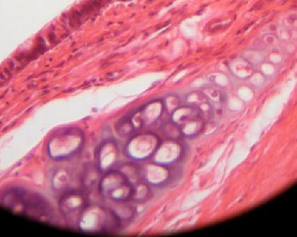
There are two other important types of epithelia:Pseudostratified epithelium and Transitional epithelium
Pseudostratified Epithelium |
Transitional Epithelium |
| Pseudostratified epithelium is really a simple epithelium (it is only one cell layer thick). All of the cells extend between the apical surface and the basal surface, yet many cells have only a tiny bit reaching to the free surface or the basal surface. Pseudostratified epithelium appears stratified (thus the name--"false stratified") because the nuclei are placed at different levels and when viewed under a microscope the tissue appears to be multiple layers thick. This tissue is often covered in cilia, which move fluids (e.g., in the trachea). | Transitional epithelium is really a form of stratified epithelium. It is different than other stratified epithelium because it allows considerable stretch (it is found in bladder, ureters and the calyces of the kidney which fill with urine and expand). The surface cells of transitional epithelium appear bulbous and misshapen when the organ is not being streched, but they become flat and thin when the organ is stretched. We will examine this tissue in the unstretched form. |
|
|

|

|
 |
Try to identify the major epithelium on each of the slides below. Although the major characteristics are still there, sometimes the preparation or particular section of a slide will make it difficult to distinguish the specific type of epithelium. Rest your mouse pointer on each picture after you've attempted to find and name the epithelium to see the tissue type.

|

|

|

|
Now go to Histology Zoomer Home Page and try the Self-guided epithelium library and the Epithelium quiz
Unlike epithelium, connective tissues do not have a free surface. However, on a microscope slide you may see open spaces around the connective tissues.
Connective tissue generally consists of a considerable amount of extracellular material called matrix. Usually there is more extracellular matrix than cellular material. The matrix is composed of varying types of fluids and proteins and fibers.
Connective tissue can be separated into loose and dense connective tissue, cartilage, bone, blood, and adipose.
This type of connective tissue cushions organs and underlies epithelia. It is characterized by having cells that produce fibers (fibroblasts) and macrophages as well as collagen, reticular, and elastic fibers in the matrix.
| Image from Areolar Slide | Areolar Connective Tissue under the epithelium of the epidermis in skin |
 |

|
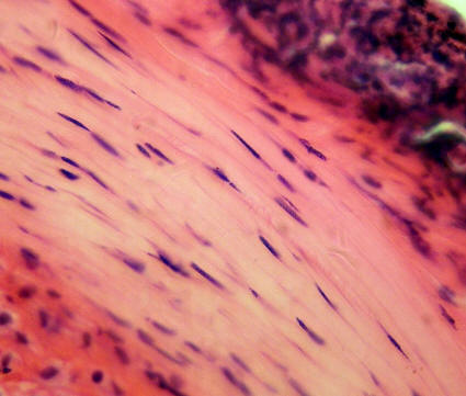
There are two types of dense connective tissue proper. They are dense regular connective tissue and dense irregular connective tissue. Both types contain an extensive matrix full of collagen fibers. These fibers are produced by fibroblasts in the tissue. The major difference between the two tissues, besides where they are found, is the direction in which the fibers run.
| Dense Regular Connective Tissue -- Found in tendons and ligaments and it is dense with collagen fibers that run parallel to one another (thus the name). This design allows the tissue to resist tension pulling in one direction. | Dense Irregular Connective Tissue -- Found in joint capsules, the dermis of the skin and in hollow organs on the digestive tract. It is dense with collagen fibers that run in many different directions. This design allows the tissue to resist tension pulling from numerous directions. |
 |

|

|
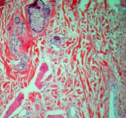
Cartilage is a form of connective tissue that has an extensive matrix surrounding the cartilage producing cells (chondroblasts or chondrocytes). These cells are trapped inside of small spaces called lacunae (little lakes).The major difference in identifying the different cartilage types is found in the visible components of the matrix. Hyaline cartilage lacks any visible fibers in the matrix, where elastic cartilage looks similar to hyaline cartilage, but it has elastic fibers in the matrix. Fibrocartilage is very dense with thick collagen fibers throughout the matrix.
Hyaline Cartilage |
Elastic Cartilage |
Fibrocartilage |

|

|

|

|

|

|
Bone is perhaps one of the easiest connective tissues to identify. The matrix in bone is made up of calcium salts that have been deposited on collagen fibers. The result of this deposition in dense bone is the formation of layers of matrix often in concentric rings (part of a structure called the osteon). Like cartilage, bone has lacunae. In bone, however, the lacunae (note the small dark structures in the second slide at high magnification) contain osteocytes .

|

|
These are two more very recognizable tissues.
It would seem that blood should not be connective tissue, but it has the same characteristics as the other types of connective tissue: it comes from the same embryonic tissue and it contains considerable extracellular matrix (blood plasma). The most abundant cells in blood are erythrocytes (red blood cells) and the darker cells on the slide are leukocytes (white blood cells).
Adipose is the tissue that stores lipids. It is what we commonly call fat. It is characterized by large cells (adipocytes) that are filled with lipids. Note how the nucleus is pushed to the edge of each cell.
| Blood | Adipose |
|
|

|

|

|
Take a quick Connective Tissue Quiz below. Try to identify the major type of connective tissue in the picture on the left, then move your mouse over the answer link on the right to see the name for the tissue.
Now go to Histology Zoomer Home Page and try the Self-guided connective tissue library and the connective tissue quiz
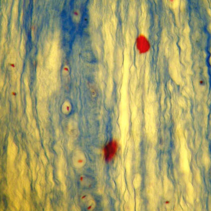
Nervous tissue is composed of neurons and their supporting cells (glial cells or neuroglia).
| Below are two images from neuron smear slides. The image on the left is at low power and the one on the right at high power. Note the large dark cells with long projections sticking out of them. These are the neurons. Note the smaller cells (you can easily see the nuclei) on the image on the right. These are glial cells. | |

|

|
| Below are cross-sections through a peripheral nerve take from an "Artery, Vein and Nerve" slide. A nerve is basically a bundle of axons (long projections) from various neurons. Around these axons (note the dark centers in the middle of the circular structures on the high power image on the right) are glial cells called Schwann cells. These make up the white myelin which extends around the each axon. | |
|
|
 |
Now go to Histology Zoomer Home Page and try the Self-guided Nervous tissue library and the Nervous tissue quiz
Muscle tissue is excitable and contractile (it shortens forcefully). The three types of muscle tissues are skeletal muscle, cardiac muscle and smooth muscle.
| Skeletal Muscle: Note that the cells (running vertically) are long, straight, straited and multinucleated. | Cardiac Muscle: Note that the cells are much more branching than skeletal muscle, but are still striated. The key feature is the dark lines that run across the tissue between cells. These are intercalated discs and are only found in cardiac muscle. | Smooth Muscle: Note that striations are not present (thus the name) and that the cells are flattened and stretched out (spindle shaped). |
 |

|

|
Now go to Histology Zoomer Home Page and try the Self-guided Muscle library and the Muscle quiz